
HEART ULTRASOUND
Visible heart health: precision in ultrasound
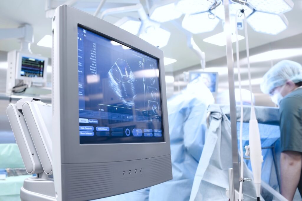
Cardiac ultrasound, technically known as echocardiography, is the window to your heart. Without harmful radiation exposure, it enables PD Dr. med. Raphael Bruno, to see your heart in motion and in real time. We assess the structure of your heart valves, the thickness of the heart muscle and the exact pumping function. Using the latest high-end technology at the Neue Stahlhof, we make the smallest changes visible long before they become a problem.
HEART ULTRASOUND
Clarity through sound:
Technology is the tool, experience is the key. PD Dr. med. Raphael Bruno uses the latest generation of high-resolution ultrasound systems to create a dynamic image of your heart. We analyze not only the anatomy, but the entire mechanics of your cardiovascular system to offer you maximum safety.
When is an ultrasound examination of the heart advisable?
This examination is the most important tool in basic cardiologic diagnostics and the first choice for almost every question.
For shortness of breath & tirednessTo reliably rule out or prove the cause of heart failure (cardiac insufficiency).
For heart murmursFor precise clarification of valve changes heard by the family doctor.
For high blood pressure patientsTo determine whether the heart is already suffering from the high pressure (muscle thickening).
In the sports check-upTo exclude congenital heart defects or wall thickening before intensive exercise.
What we measure - details of ultrasound diagnostics
In the New Stahlhof we use specialized software analyses that go far beyond the standard ultrasound image. This provides us with objective data for your individual therapy.
| Field of investigation | Method & medical benefits |
| Pumping capacity | LVEF determinationMeasurement of the percentage of blood ejected per beat to assess the strength of the heart. |
| Tissue stretching | Speckle Tracking (Strain)Highly sensitive measurement of muscle fiber movement for early detection of the smallest damage. |
| Flow velocities | CW & PW DopplerMeasurement of the pressure gradients across the flaps for exact classification of faults. |
| Pericardium check | Pericardial scanExclusion of fluid accumulation (effusion) or inflammation of the pericardium. |
| Atrial dimensions | LA volume indexDetermination of atrial size as an important risk factor for atrial fibrillation. |
| Right heart function | TAPSE measurementSpecial analysis of the right ventricle to assess the pulmonary circulation. |
HEART ULTRASOUND
Cardiac ultrasound: Frequently asked questions
Early clarification of symptoms or risk factors is the key to long-term heart health. Our cardiology practice in Düsseldorf offers you the right specialized care for every need.
A thorough examination with PD Dr. Bruno takes about 15 to 20 minutes. Including the discussion of the results, you should allow about 30 minutes.
Not directly. But we see the Consequences of blockages, such as wall movement disorders. For a direct view of the vessels, we use cardiac CT or the catheter.
Occasionally, Dr. Bruno will ask you to hold your breath for a moment or turn to the left side to optimize the image quality.
The examination is completely painless. Only the slight pressure of the ultrasound probe on the chest may be felt for a short time.
HEART ULTRASOUND
Cardiac medicine to watch in the Stahlhof
In our practice in the New Stahlhof we want you to understand how your heart works. During the examination by PD Dr. med. Raphael Bruno you can follow the images of your beating heart live on a separate monitor. In a relaxed atmosphere and without time pressure, Dr. Bruno will explain the findings to you step by step. We combine high-end diagnostics with personal attention so that you can feel completely safe.